Back Of Neck Anatomy Muscles - Human Anatomy Showing Deep Muscles In The Neck And Upper Back Art Print Art Com. The superficial group acts on upper limbs and. Muscles that act on the back. Neck muscles help support the cervical spine and contribute to movements of the head, neck, upper back, and posterior longitudinal ligament (pll). The neck muscles (and neck anatomy on the whole) are responsible for head movement, stabilizing the upper region of the body, assisting in the neck muscles include the scalenes, which attach the cervical vertebrae to the thoracic cage, and the sternocleidomastoid, which attaches the skull to the. Many in the neck help to stabilize or move the head.
There are several individual muscles within the back anatomy, and it's important to take a quick look the image below to shows all the major back muscles (as well as some neck muscles) Superficial muscles are the muscles closest to the skin surface and can usually be seen while a body is performing actions. Intermediate layer of back muscles. William is a final year medical student in australia who has taught anatomy to tertiary science and medical students since 2010. The head rests on the top part of the vertebral column, with the skull joining at c1.
The Back Of Head Neck Muscles Anatomy Page 1 Line 17qq Com from img.17qq.com Learn anatomy faster and remember everything you learn. Muscle attached to the mastoid and the. The back muscles stabilize and move the vertebral. Week 2 anatomy (back/neck muscles). Watch cervical muscle anatomy animation. William is a final year medical student in australia who has taught anatomy to tertiary science and medical students since 2010. Here the extrinsic back muscles are classified into logical subgroups to facilitate knowledge. Muscles that act on the back.
Many in the neck help to stabilize or move the head.
This article describes the anatomy of the head and neck of the human body, including the brain, bones, muscles, blood vessels, nerves, glands, nose, mouth, teeth, tongue, and throat. This is a table of skeletal muscles of the human anatomy. Almost every muscle constitutes one part of a pair of identical bilateral muscles, found on both sides, resulting in approximately 320 pairs of muscles. Muscles that act on the back. The back muscles stabilize and move the vertebral column, and are grouped according to the lengths and direction of the fascicles. The deep back muscles lie immediately adjacent to the vertebral column and ribs. The three scalene muscles are found forming the floor of the posterior triangle. The splenius capitis and cervicis (spinotransversales muscles). There are around 650 skeletal muscles within the typical human body. Human muscle system, the muscles of the human body that work the skeletal system, that are under voluntary control, and that the following sections provide a basic framework for the understanding of gross human muscular anatomy, with descriptions of the large muscle groups and their actions. Head and neck anatomy is important when considering pathology affecting the same area. We will attempt to provide a simplified overview of this complex anatomy. The muscle is a thick long cord with two heads on the bias coming from the mastoid process through the neck to grudinoklyuchichnomu articulation.
Almost every muscle constitutes one part of a pair of identical bilateral muscles, found on both sides, resulting in approximately 320 pairs of muscles. This article describes the anatomy of the head and neck of the human body, including the brain, bones, muscles, blood vessels, nerves, glands, nose, mouth, teeth, tongue, and throat. The three scalene muscles are found forming the floor of the posterior triangle. There are four pairs of muscles that are responsible for chewing movements or mastication. Several other muscles of the back also extend up to the neck region and are partly connected with the cervical part of the vertebral column, including the trapezius, levator scapulae, splenius, iliocostalis, longissimus, rotatores, semispinalis, interspinales, and intertransversarii muscles.
Back Muscles And Low Back Pain from embed.widencdn.net Several other muscles of the back also extend up to the neck region and are partly connected with the cervical part of the vertebral column, including the trapezius, levator scapulae, splenius, iliocostalis, longissimus, rotatores, semispinalis, interspinales, and intertransversarii muscles. There are several individual muscles within the back anatomy, and it's important to take a quick look the image below to shows all the major back muscles (as well as some neck muscles) Muscle attached to the mastoid and the. Working in pairs on the left and. Almost every muscle constitutes one part of a pair of identical bilateral muscles, found on both sides, resulting in approximately 320 pairs of muscles. The posterior muscles of the neck are primarily concerned with head movements, like extension. Week 2 anatomy (back/neck muscles). The anterior and middle scalenes originate from the transverse processes of certain cervical vertebrae and attach to the first rib.
Intermediate layer of back muscles.
The suprahyoid muscles originate from the posterior muscles of the neck are primarily concerned with head movements, like extension. Related posts of anatomy of neck muscles. Almost every muscle constitutes one part of a pair of identical bilateral muscles, found on both sides, resulting in approximately 320 pairs of muscles. Neck muscles help support the cervical spine and contribute to movements of the head, neck, upper back, and posterior longitudinal ligament (pll). This is a table of skeletal muscles of the human anatomy. In this section, learn more about the anatomy of the muscles of the neck. The back muscles stabilize and move the vertebral column, and are grouped according to the lengths and direction of the fascicles. The muscles of the back that work together to support the spine, help keep the body upright and allow twist and bend in many directions. Anterior muscles of the neck. Intermediate layer of back muscles. Anatomy human body organs female. They are divided into three groups, as shown below. The posterior muscles of the neck are primarily concerned with head movements, like extension.
Working in pairs on the left and. Digastric, mylohyoid, geniohyoid, stylohyoid infrahyoid muscles: The back muscles stabilize and move the vertebral. Superficial muscles are the muscles closest to the skin surface and can usually be seen while a body is performing actions. Cervical spine anatomy is quite complex.
Pin On Dental School from i.pinimg.com The anterior and middle scalenes originate from the transverse processes of certain cervical vertebrae and attach to the first rib. This is a table of skeletal muscles of the human anatomy. The muscle is a thick long cord with two heads on the bias coming from the mastoid process through the neck to grudinoklyuchichnomu articulation. Sternohyoid, sternothyroid, thyrohyoid, omohyoid anterior vertebral muscles: Neck muscles help support the cervical spine and contribute to movements of the head, neck, upper back, and posterior longitudinal ligament (pll). The back anatomy includes the latissimus dorsi, trapezius, erector spinae, rhomboid, and the teres major. Intermediate back muscles and c. Cervical spine anatomy is quite complex.
Cervical spine anatomy is quite complex.
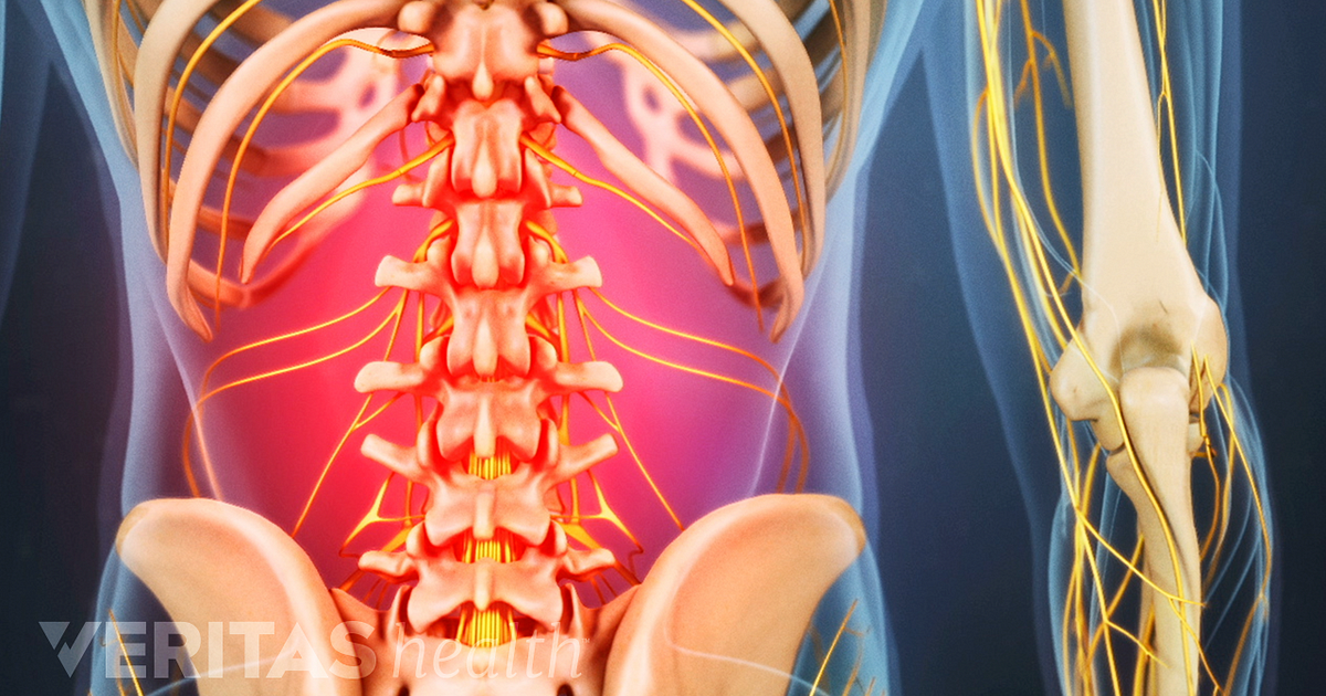
The neck muscles (and neck anatomy on the whole) are responsible for head movement, stabilizing the upper region of the body, assisting in the neck muscles include the scalenes, which attach the cervical vertebrae to the thoracic cage, and the sternocleidomastoid, which attaches the skull to the. Head and neck anatomy is important when considering pathology affecting the same area. The back muscles can be three types. The major muscle of the back of the neck, the trapezius, is involved in movements of the scapula and is dealt with in the next section, on the muscles in this view of a male figure with one arm up and one arm on the hip, there is a tremendous number of clearly defined anatomical shapes, large and small. Learn anatomy faster and remember everything you learn. The superficial group acts on upper limbs and. Anatomy human body organs female. The back muscles stabilize and move the vertebral column, and are grouped according to the lengths and direction of the fascicles. The head rests on the top part of the vertebral column, with the skull joining at c1. Neck muscles help support the cervical spine and contribute to movements of the head, neck, upper back, and posterior longitudinal ligament (pll). Watch cervical muscle anatomy animation. The pll starts at c2 and goes down the back of the vertebral bodies and intervertebral discs. The extensors and rotators of the head and neck:
Sternohyoid, sternothyroid, thyrohyoid, omohyoid anterior vertebral muscles: back of neck anatomy. The muscles of the anterior neck assist in deglutition (swallowing) and speech by controlling the positions of the larynx (voice box), and the hyoid bone, a the back muscles stabilize and move the vertebral column, and are grouped according to the lengths and direction of the fascicles.
Share :
Post a Comment
for "Back Of Neck Anatomy Muscles - Human Anatomy Showing Deep Muscles In The Neck And Upper Back Art Print Art Com"


Post a Comment for "Back Of Neck Anatomy Muscles - Human Anatomy Showing Deep Muscles In The Neck And Upper Back Art Print Art Com"